
On 5 October 2026 the journal BMC Medical Education published, as an early version ahead of its final record, a study by six authors from Shantou University Medical College in China — Zhijin Lei, Junwei Chen, Bin Zhang, Shukun Zhao, Zexin Xu and Runhua Lin — titled “Integrating autopsy-based digital case resources into undergraduate pathology teaching: a two-cohort controlled study in cardiovascular pathology”. The undergraduate cardiovascular pathology module was taught to one group with a model built on autopsy-derived digital case resources, and to a control group with conventional lecture-based teaching supported by static pathological images and routine slide observation.
The comparison was run in two independent cohorts. In cohort 1, with 87 students in each group, the mean stage assessment score was 85.40 against 78.05 (P = 0.014, Cohen’s d = 0.38). In cohort 2, with 134 students in each group, it was 86.64 against 79.48 (P = 0.003, d = 0.37). Pooled, the figures were 86.15 against 78.91 (P < 0.001, d = 0.37), and the group-by-cohort interaction was not significant (P = 0.960) — no evidence that the difference changed from one cohort to the next.
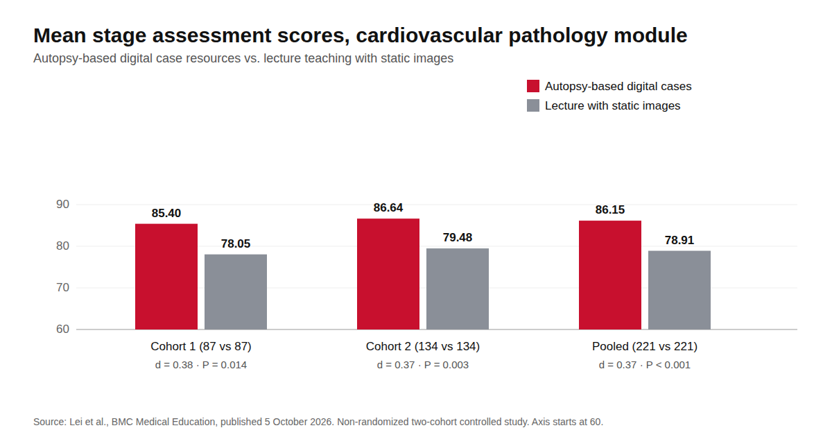
Figures from the study in BMC Medical Education (2026). © Vardix Group
On 24 September we wrote about a case-based redesign of a pathology module at Peking University, where scores rose in a class of 54 students but there was no control group, and the authors said a controlled study was still needed. The Shantou study is not that follow-up — it is a different university and a different module — but it has the design that piece was missing: a control group, and a second cohort.
What changed in the classroom
The intervention was a four-step model: case introduction, lesion observation and analysis, clinicopathological correlation, and summary and reflection. The autopsy-based digital case resources were used before, during and after class. In an anonymous post-course questionnaire, which only the intervention group completed, the domains rated highest in both cohorts were clinicopathological correlation, diagnostic and differential reasoning, and understanding of disease processes.
What the study does not show
The authors are direct about the limit: the study is not randomized, and they write that causal effects cannot be established from it. The model was associated with higher scores; it was not shown to cause them. The questionnaire describes how the intervention group saw the course and has no control-group counterpart to compare with. The version published on 5 October is an early one, which the journal says may still be edited before the final record.
It also says nothing about 3D. The case resources in Shantou were derived from autopsies; the study does not test any 3D model or simulator, and we do not read it as evidence for one.
Where Pathology3D fits
What the study does support is the structure: a course that puts a case and its lesions in front of the student, and then asks for the link between morphology and the clinical picture. That needs case material, and a department needs a steady supply of it. On 3 October we wrote about one hospital’s autopsy numbers — at that hospital, autopsies fell from 3.9% to 0.57% of inpatient deaths over 2019–2025.
Pathology3D, which VARDIX distributes to medical and pharmacy universities, is a 3D screen simulator for general and special pathological anatomy. A student selects a disease or syndrome, loads the target 3D organ and models the pathomorphological changes stage by stage at the macro level, then matches the case against photographs of histological slides at the micro level. A results table analyses the student’s answers afterwards. It runs on Windows, Linux and macOS.
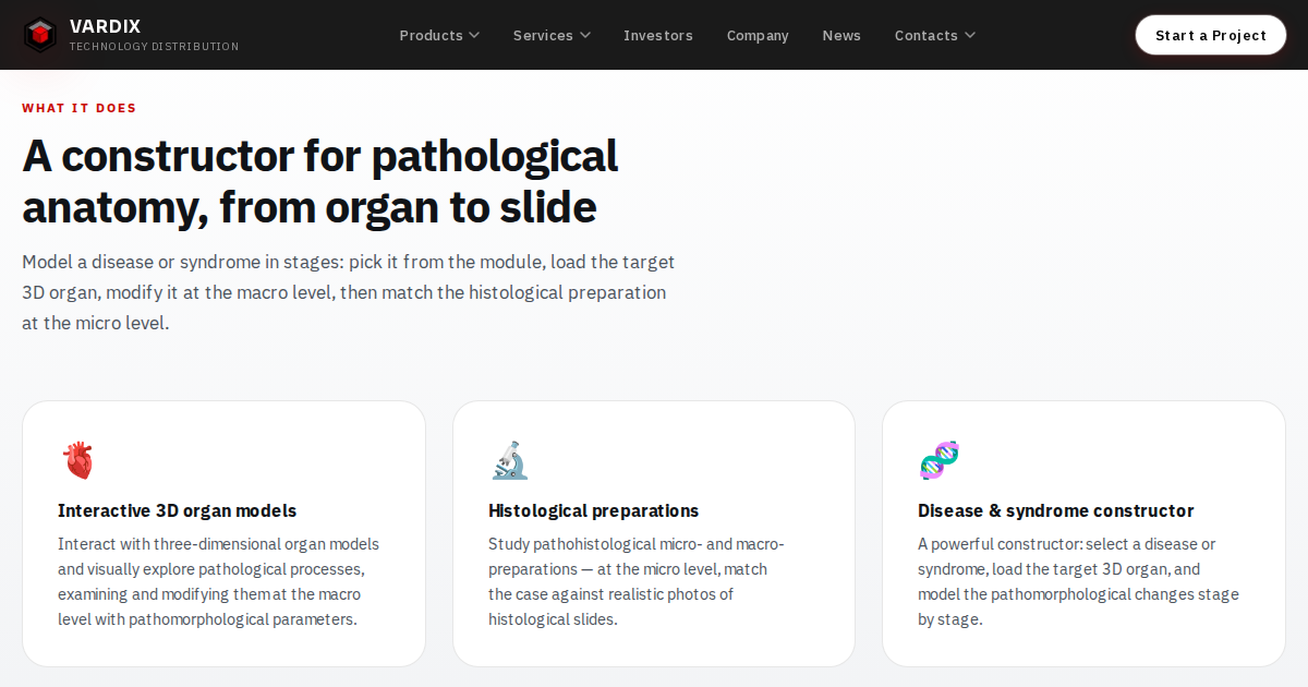
Pathology3D: from the 3D organ to the histological slide. © Vardix Group
Our reading, not the study’s finding: in a module built like the Shantou one, Pathology3D can supply the lesion-observation step — the organ at a given stage of a disease, then the slide — when the course needs it rather than when an archived case happens to fit. It does not replace an autopsy archive, and it does not teach clinicopathological reasoning on its own; that is the work of the course around it.
For distributors
The buyers are departments of pathological anatomy at medical and pharmacy universities. The conversation with them does not need a claim that 3D outperforms anything — we know of no study showing that. It needs the point this one makes: case-based pathology teaching now has a controlled comparison across two cohorts behind it, and a case-based course has to get its organs from somewhere. If your territory includes medical schools, we would like to talk.